
Helmholtz Diabetes Center Institut für Pankreatische Inselzellforschung
Die Mission des IPI ist der Schutz und die Wiederherstellung der Insulin-produzierenden Betazellen der Bauchspeicheldrüse zur Prävention und Heilung des Diabetes mellitus.
Die Mission des IPI ist der Schutz und die Wiederherstellung der Insulin-produzierenden Betazellen der Bauchspeicheldrüse zur Prävention und Heilung des Diabetes mellitus.
Über uns
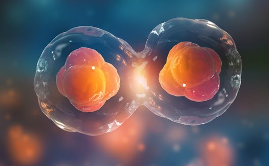
Die Forschung am IPI konzentriert sich auf die Langerhans´schen Inseln der Bauchspeicheldrüse. In diesen Inseln befinden sich die Insulin-produzierenden Betazellen welche beim Typ-1 und Typ-2 Diabetes geschädigt oder gar zerstört werden. Das Studium der zugrunde liegenden Mechanismen und deren besseres Verständnis helfen den Wissenschaftlern bei der Entwicklung neuer Therapieansätze.
Seit Januar 2016 gehört das IPI als Satelliten-Institut zu Helmholtz Munich und bildet den Kern des bereits zuvor gegründeten Paul-Langerhans-Instituts Dresden (PLID) des Deutschen Zentrums für Diabetesforschung (DZD e.V.).
Derzeit umfasst das IPI 16 Forschungsgruppen, die sich mit verschiedenen Aspekten der Diabeteserkrankung beschäftigen. Die Wissenschaftler arbeiten daran, die Mechanismen zu entschlüsseln, die die Zerstörung und/oder Funktionseinschränkung der Betazellen bedingen und versuchen darüber hinaus, neue Ansätze zu entwickeln um die geschädigten bzw. zerstörten Betazellen zu ersetzen.
Unsere Mission ist daher der Schutz und die Wiederherstellung der Insulin-produzierenden Betazellen der Bauchspeicheldrüse zur Prävention und Heilung des Diabetes mellitus.
Ein internationales Wissenschaftlerteam verschiedenster Fachrichtungen konnte über die letzten Jahre für den Standort Dresden gewonnen werden und macht das IPI zu einem der führende Orte für Diabetesforschung in Deutschland. Die interdisziplinäre Zusammenarbeit und die enge Verknüpfung von Experten verschiedener Fachdisziplinen wie der Genetik, Immunologie, Zell- und Entwicklungsbiologie mit den klinischen Abteilungen der Inneren Medizin und der Viszeral-, Thorax- und Gefäßchirurgie oder den Stammzellexperten garantieren dabei eine translationale Ausrichtung der Forschung. Die exzellente Forschungsinfrastruktur am Dresdner Standort stellt die Basis für zukünftige wissenschaftliche Spitzenleistungen.


 micrograph of a beta cell of a islet of Langerhans showing the typical insulin granules (blue), mitochondria (green) and nucleus (purple).")
![[Translate to German:]](https://images.admiralcloud.com/v3/deliverEmbed/83895c99-5670-4e4c-bca2-1fc6e0d959dd/image/cropperjsfocus/915/550/0,0,960,469,0,1,1/480,234.5?poc=true "[Translate to German:]")
![[Translate to German:]](https://images.admiralcloud.com/v3/deliverEmbed/ac4981a4-3b07-42c6-a839-d6b7337b402e/image/cropperjsfocus/915/550/0,0,1920,1457,0,1,1/960,728.5?poc=true "[Translate to German:]")
![[Translate to German:]](https://images.admiralcloud.com/v3/deliverEmbed/811349a6-9231-4ffe-8cb0-a646341bef7b/image/cropperjsfocus/915/550/0,0,960,560,0,1,1/479.99999999999994,280?poc=true "[Translate to German:]")

![[Translate to German:]](https://images.admiralcloud.com/v3/deliverEmbed/b46e5b15-e227-411a-9643-4f333e6029e3/image/cropperjsfocus/915/550/0,0,1280,768,0,1,1/640,384?poc=true "[Translate to German:]")


Unsere Mitarbeiter am IPI


Publikationen
Weiterlesen2024 Wissenschaftlicher Artikel in Cell Reports
Impaired islet function and normal exocrine enzyme secretion occur with low inter-regional variation in type 1 diabetes.
Histopathological heterogeneity in the human pancreas is well documented; however, functional evidence at the tissue level is scarce. Herein, we investigate in situ glucose-stimulated islet and carbachol-stimulated acinar cell secretion across the pancreas head (PH), body (PB), and tail (PT) regions in donors without diabetes (ND; n = 15), positive for one islet autoantibody (1AAb+; n = 7), and with type 1 diabetes (T1D; <14 months duration, n = 5). Insulin, glucagon, pancreatic amylase, lipase, and trypsinogen secretion along with 3D tissue morphometrical features are comparable across regions in ND. In T1D, insulin secretion and beta-cell volume are significantly reduced within all regions, while glucagon and enzymes are unaltered. Beta-cell volume is lower despite normal insulin secretion in 1AAb+, resulting in increased volume-adjusted insulin secretion versus ND. Islet and acinar cell secretion in 1AAb+ are consistent across the PH, PB, and PT. This study supports low inter-regional variation in pancreas slice function and, potentially, increased metabolic demand in 1AAb+.
2024 Nature metabolism
Author Correction: Every islet matters: Improving the impact of human islet research.
In the version of this article initially published, the name of the Human Pancreas Analysis Program was written incorrectly and has now been amended in the HTML and PDF versions of the article.
2024 Wissenschaftlicher Artikel in Biological Procedures Online
Fast track adaptation of oncolytic coxsackie B3 virus to resistant colorectal cancer ells - a method to personalize virotherapy.
BACKGROUND: The efficacy of oncolytic viruses (OV) in cancer treatment depends on their ability to successfully infect and destroy tumor cells. However, patients' tumors vary, and in the case of individual insensitivity to an OV, therapeutic efficacy is limited. Here, we present a protocol for rapid generation of tumor cell-specific adapted oncolytic coxsackievirus B3 (CVB3) with enhanced oncolytic potential and a satisfactory safety profile. This is achieved by combining directed viral evolution (DVE) with genetic modification of the viral genome and the use of a microRNA-dependent regulatory tool. METHODS: The oncolytic CVB3 variant PD-H was adapted to the refractory colorectal carcinoma cell line Colo320 through serial passaging. XTT assays and virus plaque assays were used to determine virus cytotoxicity and virus replication in vitro. Recombinant PD-H variants were generated through virus mutagenesis. Apoptosis was detected by Western blots, Caspase 3/7 assays, and DAPI staining. The therapeutic efficacy and safety of the adapted recombinant OV PD-SK-375TS were assessed in vivo using a subcutaneous Colo320 xenograft mouse model. RESULTS: PD-H was adapted to the colorectal cancer cell line Colo320 within 10 passages. Sequencing of passage 10 virus P-10 revealed a heterogenous virus population with five nucleotide mutations resulting in amino acid substitutions. The genotypically homogeneous OV PD-SK was generated by inserting the five detected mutations of P-10 into the genome of PD-H. PD-SK showed significantly stronger replication and cytotoxicity than PD-H in Colo320 cells, but not in other colorectal carcinoma cell lines. Increase of apoptosis induction was detected as key mechanisms of Colo320 cell-specific adaptation of PD-SK. For in vivo safety PD-SK was engineered with target sites of the miR-375 (miR-375TS) to exclude virus replication in normal tissues. PD-SK-375TS, unlike the PD-H-375TS not adapted homolog suppressed the growth of subcutaneous Colo320 tumors in nude mice without causing any side effects. CONCLUSION: Taken together, here we present an optimized protocol for the rapid generation of tumor cell-specific adapted oncolytic CVB3 based on the oncolytic CVB3 strain PD-H. The protocol is promising for the generation of personalized OV for tumor therapy and has the potential to be applied to other OV.
2024 Wissenschaftlicher Artikel in Cell Reports
OSBP-mediated PI(4)P-cholesterol exchange at endoplasmic reticulum-secretory granule contact sites controls insulin secretion.
Insulin is packaged into secretory granules that depart the Golgi and undergo a maturation process that involves changes in the protein and lipid composition of the granules. Here, we show that insulin secretory granules form physical contacts with the endoplasmic reticulum and that the lipid exchange protein oxysterol-binding protein (OSBP) is recruited to these sites in a Ca2+-dependent manner. OSBP binding to insulin granules is positively regulated by phosphatidylinositol-4 (PI4)-kinases and negatively regulated by the PI4 phosphate (PI(4)P) phosphatase Sac2. Loss of Sac2 results in excess accumulation of cholesterol on insulin granules that is normalized when OSBP expression is reduced, and both acute inhibition and small interfering RNA (siRNA)-mediated knockdown of OSBP suppress glucose-stimulated insulin secretion without affecting insulin production or intracellular Ca2+ signaling. In conclusion, we show that lipid exchange at endoplasmic reticulum (ER)-granule contact sites is involved in the exocytic process and propose that these contacts act as reaction centers with multimodal functions during insulin granule maturation.
2024 Wissenschaftlicher Artikel in Science Advances
Coxsackievirus infection induces direct pancreatic β cell killing but poor antiviral CD8+ T cell responses.
Coxsackievirus B (CVB) infection of pancreatic β cells is associated with β cell autoimmunity and type 1 diabetes. We investigated how CVB affects human β cells and anti-CVB T cell responses. β cells were efficiently infected by CVB in vitro, down-regulated human leukocyte antigen (HLA) class I, and presented few, selected HLA-bound viral peptides. Circulating CD8+ T cells from CVB-seropositive individuals recognized a fraction of these peptides; only another subfraction was targeted by effector/memory T cells that expressed exhaustion marker PD-1. T cells recognizing a CVB epitope cross-reacted with β cell antigen GAD. Infected β cells, which formed filopodia to propagate infection, were more efficiently killed by CVB than by CVB-reactive T cells. Our in vitro and ex vivo data highlight limited CD8+ T cell responses to CVB, supporting the rationale for CVB vaccination trials for type 1 diabetes prevention. CD8+ T cells recognizing structural and nonstructural CVB epitopes provide biomarkers to differentially follow response to infection and vaccination.
2024 Review in Nature Protocols
Modular segmentation, spatial analysis and visualization of volume electron microscopy datasets.
Volume electron microscopy is the method of choice for the in situ interrogation of cellular ultrastructure at the nanometer scale, and with the increase in large raw image datasets generated, improving computational strategies for image segmentation and spatial analysis is necessary. Here we describe a practical and annotation-efficient pipeline for organelle-specific segmentation, spatial analysis and visualization of large volume electron microscopy datasets using freely available, user-friendly software tools that can be run on a single standard workstation. The procedures are aimed at researchers in the life sciences with modest computational expertise, who use volume electron microscopy and need to generate three-dimensional (3D) segmentation labels for different types of cell organelles while minimizing manual annotation efforts, to analyze the spatial interactions between organelle instances and to visualize the 3D segmentation results. We provide detailed guidelines for choosing well-suited segmentation tools for specific cell organelles, and to bridge compatibility issues between freely available open-source tools, we distribute the critical steps as easily installable Album solutions for deep learning segmentation, spatial analysis and 3D rendering. Our detailed description can serve as a reference for similar projects requiring particular strategies for single- or multiple-organelle analysis, which can be achieved with computational resources commonly available to single-user setups.
2024 Wissenschaftlicher Artikel in Cell Reports
Purification of time-resolved insulin granules reveals proteomic and lipidomic changes during granule aging.
Endocrine cells employ regulated exocytosis of secretory granules to secrete hormones and neurotransmitters. Secretory granule exocytosis depends on spatiotemporal variables such as proximity to the plasma membrane and age, with newly generated granules being preferentially released. Despite recent advances, we lack a comprehensive view of the molecular composition of insulin granules and associated changes over their lifetime. Here, we report a strategy for the purification of insulin secretory granules of distinct age from insulinoma INS-1 cells. Tagging the granule-resident protein phogrin with a cleavable CLIP tag, we obtain intact fractions of age-distinct granules for proteomic and lipidomic analyses. We find that the lipid composition changes over time, along with the physical properties of the membrane, and that kinesin-1 heavy chain (KIF5b) as well as Ras-related protein 3a (RAB3a) associate preferentially with younger granules. Further, we identify the Rho GTPase-activating protein (ARHGAP1) as a cytosolic factor associated with insulin granules.
2024 Review in Diabetes
Bridging the Gap: Pancreas tissue slices from organ and tissue donors for the study of diabetes pathogenesis.
UNLABELLED: Over the last two decades, increased availability of human pancreatic tissues has allowed for major expansions in our understanding of islet biology in health and disease. Indeed, studies of fixed and frozen pancreatic tissues, as well as efforts using viable isolated islets obtained from organ donors, have provided significant insights toward our understanding of diabetes. However, the procedures associated with islet isolation result in distressed cells that have been removed from any surrounding influence. The pancreas tissue slice technology was developed as an in situ approach to overcome certain limitations associated with studies on isolated islets or fixed tissue. In this Perspective, we discuss the value of this novel platform and review how pancreas tissue slices, within a short time, have been integrated in numerous studies of rodent and human islet research. We show that pancreas tissue slices allow for investigations in a less perturbed organ tissue environment, ranging from cellular processes, over peri-islet modulations, to tissue interactions. Finally, we discuss the considerations and limitations of this technology in its future applications. We believe the pancreas tissue slices will help bridge the gap between studies on isolated islets and cells to the systemic conditions by providing new insight into physiological and pathophysiological processes at the organ level. ARTICLE HIGHLIGHTS: Human pancreas tissue slices represent a novel platform to study human islet biology in close to physiological conditions. Complementary to established technologies, such as isolated islets, single cells, and histological sections, pancreas tissue slices help bridge our understanding of islet physiology and pathophysiology from single cell to intact organ. Diverse sources of viable human pancreas tissue, each with distinct characteristics to be considered, are available to use in tissue slices for the study of diabetes pathogenesis.
2024 Wissenschaftlicher Artikel in eLife
Regulation of multiple signaling pathways promotes the consistent expansion of human pancreatic progenitors in defined conditions.
The unlimited expansion of human progenitor cells in vitro could unlock many prospects for regenerative medicine. However, it remains an important challenge as it requires the decoupling of the mechanisms supporting progenitor self-renewal and expansion from those mechanisms promoting their differentiation. This study focuses on the expansion of human pluripotent stem (hPS) cell-derived pancreatic progenitors (PP) to advance novel therapies for diabetes. We obtained mechanistic insights into PP expansion requirements and identified conditions for the robust and unlimited expansion of hPS cell-derived PP cells under GMP-compliant conditions through a hypothesis-driven iterative approach. We show that the combined stimulation of specific mitogenic pathways, suppression of retinoic acid signaling, and inhibition of selected branches of the TGFβ and Wnt signaling pathways are necessary for the effective decoupling of PP proliferation from differentiation. This enabled the reproducible, 2000-fold, over 10 passages and 40-45 d, expansion of PDX1+/SOX9+/NKX6-1+ PP cells. Transcriptome analyses confirmed the stabilization of PP identity and the effective suppression of differentiation. Using these conditions, PDX1+/SOX9+/NKX6-1+ PP cells, derived from different, both XY and XX, hPS cell lines, were enriched to nearly 90% homogeneity and expanded with very similar kinetics and efficiency. Furthermore, non-expanded and expanded PP cells, from different hPS cell lines, were differentiated in microwells into homogeneous islet-like clusters (SC-islets) with very similar efficiency. These clusters contained abundant β-cells of comparable functionality as assessed by glucose-stimulated insulin secretion assays. These findings established the signaling requirements to decouple PP proliferation from differentiation and allowed the consistent expansion of hPS cell-derived PP cells. They will enable the establishment of large banks of GMP-produced PP cells derived from diverse hPS cell lines. This approach will streamline SC-islet production for further development of the differentiation process, diabetes research, personalized medicine, and cell therapies.
2023 Wissenschaftlicher Artikel in Experimental Cell Research
Mechanism of pro-MMP9 activation in co-culture of pro-inflammatory macrophages and cardiomyocytes.
OBJECTIVE: A wide range of cardiac diseases is associated with inflammation. "Inflamed" heart tissue is infiltrated with pro-inflammatory macrophages which extensively secrete matrix metalloproteinase 9 (MMP9), a regulator of extracellular matrix turnover. As MMP9 is released from macrophages in a latent form, it requires activation. The present study addresses the role of cardiomyocytes in the course of this activation process. METHODS AND RESULTS: In mono- and co-cultures of pro-inflammatory rat macrophages (bone marrow-derived and peritoneal) and cardiomyocytes (H9C2 cell line) gelatin zymography demonstrated that activated macrophages robustly secreted latent pro-MMP9, whereas cardiomyocytes could not produce the enzyme. Co-culturing of the two cell species was critical for pro-MMP9 activation and was also accompanied by processing of cardiomyocyte-secreted pro-MMP2. A cascade of pro-MMP9 activation was initiated on macrophage membrane with pro-MMP2 cleavage. Namely, pro-inflammatory macrophages expressed an active membrane type 1 MMP (MT1MMP), which activated pro-MMP2, which in turn converted pro-MMP9. Downregulation of MT1MMP in macrophages by siRNA abolished activation of both pro-MMP2 and pro-MMP9 in co-culture. In addition, both cell species secreted MMP13 as a further pro-MMP9 activator. In co-culture, activation of pro-MMP13 occurred on membranes of macrophages and was enhanced in presence of active MMP2. Using incubations with recombinant MMPs and isolated macrophage membranes, we demonstrated that while both MMP2 and MMP13 individually had the ability to activate pro-MMP9, their combined action provided a synergistic effect. CONCLUSION: Activation of pro-MMP9 in a co-culture of pro-inflammatory macrophages and cardiomyocytes was the result of a complex interaction of several MMPs on the cell membrane and in the extracellular space. Both cell types contributed critically to pro-MMP9 processing.


